문서의 이전 판입니다!
무뇌아 [Anencephaly]
신경관 결손증 (Neural Tube Defect)이란 머리에서 등뼈 끝까지 연결되는 중추신경이 있는 어느 곳이 완전히 막히지 않아서 결손이 생기는 것이다. 대표적인 신경관 결손으로는 무뇌아와 이분 척추가 있다.
임신하기 한두 달 전부터 임신 초기까지 Folic acid 보충을 하면 신경관 결손증 발생이 줄어든다는 연구가 있다.1) 이런 기형아를 낳은 산모는 다음 임신하기 전부터 엽산을 하루에 4 mg씩 보충하면 도움이 될 가능성도 있다.
무뇌아
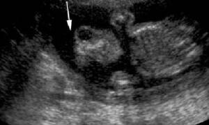
기형아의 상당 부분을 차지하며 대뇌가 없거나 거의 없는 것으로 머리뼈도 위쪽은 없다. 그냥 두면 뱃속에서 태아 시절에는 잘 죽지 않고 자라기 때문에 과거에 초음파가 없었던 시기에는 만삭까지 자라서 태어나는 아이가 많았다.
Approximately one child for every 1000 births (Central Europe). This rate varies according to populations. (Sadler, T.W. 2005)
무뇌아의 진단
요즘에는 초음파검사와 산모 혈액을 통한 기형아검사로 미리 진단하기 때문에 만삭에 태어나는 아이가 거의 없다. 임신 16-18주에 하는 산모 혈액으로 하는 기형아검사는 가능성만 제시할 뿐이고 확진은 초음파로 한다.
배 초음파로 임신 12주면 진단이 거의 100% 가능하다. 이 시기에 초음파검사를 하면 무뇌아 뿐만 아니라 세밀하지는 못해도 팔다리를 포함한 전체적인 태아 형태에 큰 이상이 없는지도 알 수 있으며 배꼽 탈장, 복벽 균열, 목뒤 임파선 비대증 등 몇 가지 중요한 기형을 발견할 수 있으므로 이 때 초음파검사를 하는 것은 상당히 가치가 있다.
기형아검사
두개골과 뇌막이 없으므로 남아 있는 뇌 부분이 양수에 노출되어 산모 혈액을 이용하는 기형아검사에서 태아 알파 단백 수치가 매우 높게 올라간다. 태아의 신체 내부가 양수에 노출됨으로 태아 신체에 많이 있으면서 기형아검사에서 검출하는 물질인 태아 알파 단백질이 양수를 통하여 산모 혈액으로 많이 들어가기 때문이다.
무뇌아의 분만
태아의 뇌가 없으면 자연적인 진통이 잘 오지 않을 뿐만 아니라 유도분만을 위해서 촉진제를 써도 정상 태아보다 진통이 잘 오지 않는다. 뇌가 없으면 부신이 발육되지 않는데, 자궁 수축에는 태아의 뇌기능이나 부신 기능이 어느 정도 필요하기 때문이다.
양수 과다증이 잘 생기는데 이때는 자궁이 너무 늘어나므로 진통이 일찍 올 때도 있지만 양막이 미리 터지고서도 진통이 잘 오지 않으면 염증이 생기지 않도록 빨리 분만해야 되는데 어려움이 따를 수 있다.
About 7% die during the pregnancy (prenatal death), 20% die during delivery; 50% have a life expectancy of between a few minutes and 1 day, 23% live more than 1 day (Jaquier 2006).
Can anencephaly be prevented?
For some time now, the aetiology of Neural Tube Defects has cited diet and environmental factors. Clinical studies have confirmed that taking a vitamin called Folic Acid reduces the risk of developing a Neural Tube Defect. . Learn more about the prevention of neural tube defects.
기타
- Botto LD et al, 1999. Neural-Tube Defects. N England J Med 341:1509-1519
- Czeizel AE, Dudas I. 1992. Prevention of first occurence of neural tube defects by periconceptional vitamin supplementation. N Engl J Med 327:1832-1835
- Holmes LB, Briscoll SG, Atkins L. 1976. Etiologie heterogeneity of neural-tube defects. N Engl J Med 1976;294:365-369
- Jaquier M, Klein A, Boltshauser E., 2006. Spontaneous pregnancy outcome after prenatal diagnosis of anencephaly, BJOG 2006; 113:951-953
- Johnson SP, Sebire NJ, Snijders RJM, Tunkel S, Nicolaides KH. Ultrasound screening for anencephaly at 10-14 weeks of gestation. Ultrasound Obstet Gynecol 1997;9:14.
- Müller F, O'Rahilly R, 1991. Development of Anencephaly and Its Variants. The American Journal of Anatomy 190:193-218 (1991)
- Sadler TW, 1998. Mechanisms of neural tube closure and defects. Ment Retard Dev Disabil Res Rev 1998;4:247-53
- Sadler TW. 2005. Embryology of Neural Tube Development. American Journal of Medical Genetics Part C 135C:2-8
- Stumpf et al. The Infant with Anencephaly, The Medical Task Force on Anencephaly, N Engl J Med 1990; 322:669-67

